Unit 1 – Sternoclavicular Joint Sprain (Preview)
Quiz: When you have completed the learning requirements for this unit, you will be able to access its quiz by clicking on the link found at the bottom of this web page. You can attempt the quiz as many times as you need to achieve a pass mark of at least 70%.
Disclaimer: Please read the Disclaimer at the bottom of this page.
Copyright © Educom Pty Ltd: All material on this website (including the text, graphics, videos and downloadable files) is owned by or licensed to Educom Pty Ltd and is subject to copyright and other intellectual property rights under international conventions.
How to Complete This Unit
This unit consists of reading material, video presentations, and practice resources. When you have completed the learning material in this unit, you will be able to access its multiple-choice quiz by clicking on the link found at the bottom of this web page. You can attempt the quiz as many times as you need to achieve a pass mark of at least 70%.
Unit Credit: 2 Hours
2 hours include allocated time for progressing through the reading material, watching the videos, considering the reflection points, final review of the unit and completing the quiz.
Content Links
The following buttons can be used to navigate the content in this unit.
Diagnostic Features
Sternoclavicular Joint Sprain
Introduction
Sternoclavicular joint injuries involve the sternoclavicular joint and its associated ligaments and can be of a traumatic or atraumatic aetiology. Traumatic injury to the sternoclavicular joint is reported to be caused most commonly by motor vehicle accidents or sporting trauma. The mechanism of injury is said to be that of compression force to the lateral shoulder, together with a vector that drives the medial end of the clavicle anteriorly or posteriorly, typically resulting in a sprain of the joint. When the injury is severe dislocation may occur. In cases of atraumatic injury, the usual mechanism is sustained activity with the arm in an elevated position.
With only half of the medial end of the clavicle in direct contact with the manubrium, the sternoclavicular joint is an inherently unstable joint relying on ligamentous support. The joint contains an intra-articular disc which acts as a shock absorber and allows for a greater range of mobility.
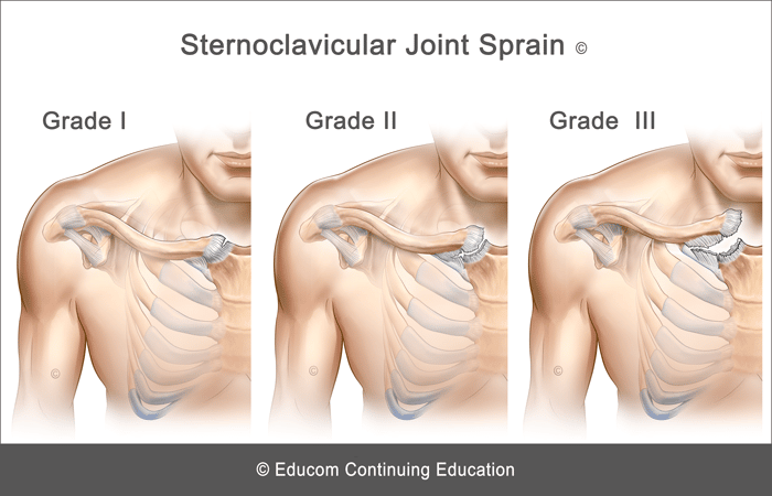
Injuries of the sternoclavicular joint are classified into three types based on their severity:
Grade I – Sprain: An incomplete tear or stretching of the sternoclavicular and costoclavicular ligaments.
Grade II – Subluxation: A complete tear of the sternoclavicular ligament but only a partial tear of the costoclavicular ligament.
Grade III – Dislocation: A complete rupture of the sternoclavicular and costoclavicular ligaments.
History and Presentation
- Anterior chest pain primarily localised to the sternoclavicular joint
- Pain aggravation with arm movement
- Pain aggravation when lying on the side of involvement
- Pain may occur with neck flexion in the supine position
- Posterior dislocation may lead to dyspnea, stridor, dysphagia, or upper limb paraesthesia
Physical Examination
- Swelling or prominence at the medial end of the clavicle
- Tenderness on palpation over the sternoclavicular joint
- May have local ecchymosis
- Pain aggravation with shoulder abduction, flexion, or horizontal adduction
- Pain aggravation with resisted neck flexion
- Shoulder special tests may aggravate pain (e.g., Painful Arc and Acromioclavicular Crossover tests)
Imaging
Plain radiography may be used to demonstrate widening of the joint or clavicular dislocation. Computerised Tomography (CT) may be required in cases of posterior dislocation to better evaluate mediastinal structures.
Red Flags
The following are examples of “red flags” for patients presenting with trauma-induced chest pain:
- History of a significant injury
- Severe pain
- Unrelenting pain
- Nocturnal pain or pain at rest
- Fever
- Deformity
- Large joint swelling
- Significant loss of range of motion
- Significant neurological impairment (suggestive of a space-occupying lesion)
- Severe tenderness on palpation or severe pain with any examination procedure
- Dyspnea, stridor, dysphagia, or upper limb paresthesia
If any “red flags” are identified during history taking and clinical examination, referral for urgent medical evaluation and further investigation is warranted.
Management
If no red flags are identified or further investigations do not reveal any contraindications to treatment, management of the condition may begin.
The management approach outlined below is based on published materials (see the References section at the end of this web page) and the clinical experience of the authors of this course. This should not be interpreted as a prescriptive guide to the treatment of this or any other condition. The use of this content is subject to the Disclaimer found at the bottom of this web page.
The following suggestions for therapeutic intervention will depend on the extent of the injury and the stage of recovery.
Conservative Therapy
- Correction of joint dysfunction with emphasis on the shoulder and spine
- Management of any associated myofascial pain syndrome
- Electrotherapy modalities
- Shoulder support (e.g., arm sling, figure-of-eight brace)
- Home advice including activity modification, rehabilitation exercises, and stretches (see below)
Home Advice
- Apply ice over the affected region for about 10 minutes, several times a day for pain control. Ice can be applied as often as every hour.
- Avoid aggravating activities.
- When pain allows, perform muscle strengthening and stretching exercises.
- When it is time to resume activities, initially perform these for only short periods.

Differential Diagnosis
Other conditions that are considered in the differential diagnosis of upper anterior chest pain include:
- Tietze Syndrome
- Sternalis Muscle Myofascial Pain Syndrome
- SCM muscles strain
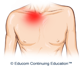
For Patients
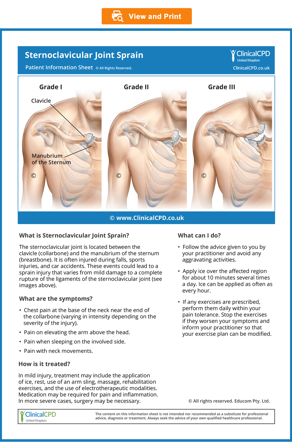
Patient Information Sheet
- Provide information to the patient about their condition to improve their understanding and enhance compliance (see the downloadable ready-to-use Patient Information Sheet below).
- In our experience, patients who are well informed about their condition are more likely to comply with the recommended management strategy, achieve good outcomes, become loyal patients, and recommend their family and friends to seek treatment.
- To view, download or print the Patient Information sheet, simply click on the image below.
© Educom Pty. Ltd. All Rights Reserved.
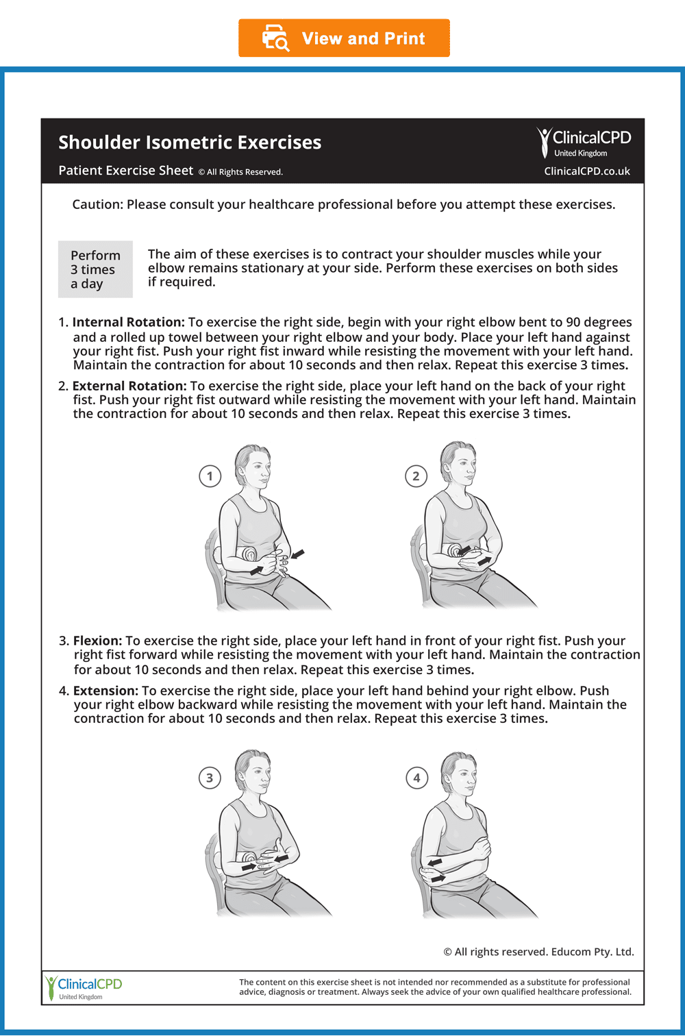
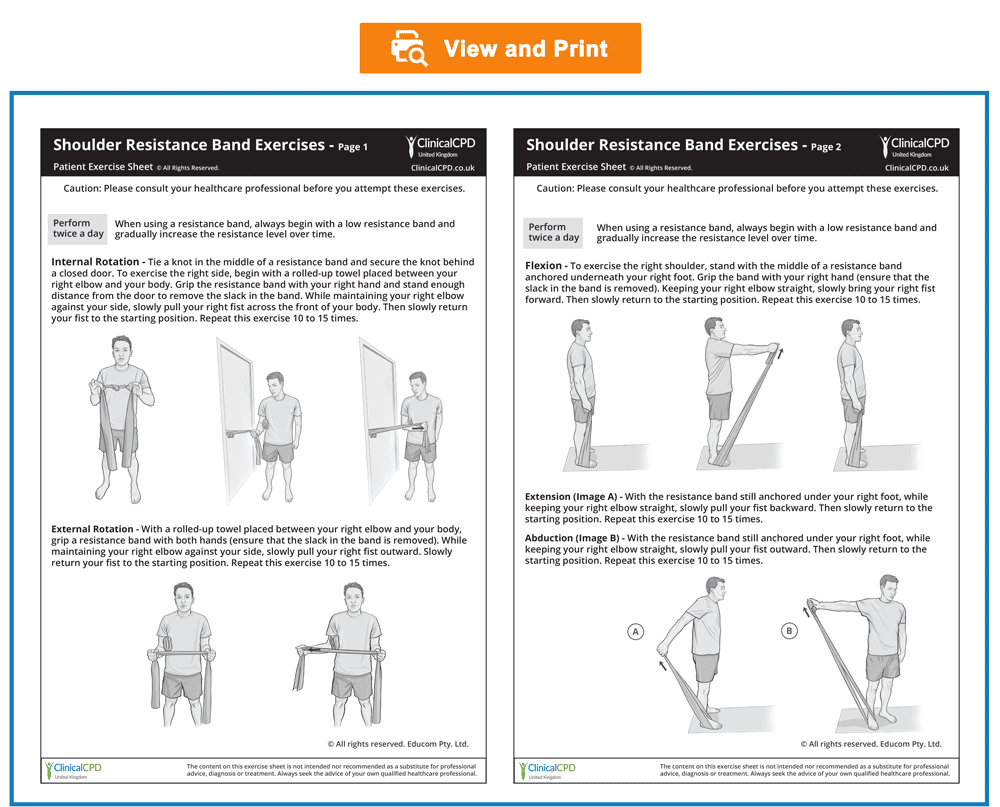
Patient Exercise Sheets
- When appropriate, the patient should gradually begin doing exercises at home.
- Always recommend warm-up activities before commencing specific exercises. Warm-up activities include simple limbering movements or prescribed strength exercises at light loads.
- Instruct your patients to perform any strengthening exercises before they perform any stretching exercises.
- Always instruct your patients to use caution when performing their rehab exercises in order to avoid overloading, overstretching, or any undue pain.
- Emphasise that they should stop any exercises that cause them concern and seek your advice at the earliest opportunity
- To view, download or print the Patient Exercise Sheet, simply click on the image below.
© Educom Pty. Ltd. All Rights Reserved.


Optimise Your Diagnostic Reasoning
Our Recommended Approach
To help you sharpen your diagnostic reasoning skills, we recommend the following steps:
- Step 1: In the following case scenario, read through the provided patient information in each section.
- Step 2: Consider the ‘Diagnostic Reasoning Question’ for that section and take some time to think about your response before you read our suggested answer.
- Step 3: To encourage critical thinking, we recommend that you first attempt to answer the question on your own. If you wish, you can hide our suggested answer by clicking on the ‘Suggested Answer’ arrow. When ready, click the arrow again to reveal our answer and compare it with your own.
Please note that the suggested answers are only indicative and not exhaustive. They serve as guidelines and should not be considered complete answers.
Patient’s History
In the sections below, you are provided with case history information. The material presents a systematic approach to performing a focused and relevant case history in order to narrow down the possible causes of the patient’s complaint. As you read the following material, you are encouraged to answer the questions posed to test your diagnostic reasoning skills.
Who is the patient and where is the pain?
When was the onset and what caused the onset?
Nick is a 23-year-old amateur wrestler who presents with acute right medial upper chest pain following an injury sustained during a wrestling match three days ago. He explains that he was thrown onto his right side, outside the wrestling area, causing his right shoulder to strike the floor. He immediately felt pain in the front of his chest and at the base of his throat. Nick points to the area over the medial end of his right clavicle as the main site of pain.
|
What differential diagnoses would you consider for this patient? |
Suggested Answer
- Clavicular fracture
- Sternoclavicular joint sprain or dislocation
- Upper rib or sternal fracture
- Muscle strain (e.g., the pectoralis major, sternocleidomastoid, or subclavius muscles)
What are the pain characteristics?
What are the aggravating and relieving factors?
What has been the course of the pain?
Nick describes his pain as a dull ache at rest, rating it 4 out of 10 in intensity. However, when he moves his right arm, the pain increases to 8 out of 10. Additionally, he experiences an increase in pain when lying on his right side or when lifting his head from a pillow while lying on his back.
He mentions that he has avoided using his right arm since the injury, and a friend with some first aid experience made him a sling, which has provided significant pain relief. Additionally, he finds that applying ice directly over the pain site offers significant, albeit temporary, relief.
|
What could be the potential causes of increased pain when Nick lies on his right side or lifts his head from a pillow while lying on his back? |
Suggested Answer
- Musculoskeletal Injury – Lying on the right side compresses injured structures (e.g., muscles, tendons, ligaments) in the shoulder, leading to increased pain. Similarly, lifting the head while lying down can engage the structures in the upper chest area, exacerbating the pain due to the added strain on the affected tissues (e.g., clavicle fracture or sternoclavicular joint injury).
- Referred Pain – The pain could be referred from another area, such as the neck or upper thoracic spine, which might explain why certain positions aggravate the symptoms.
- Inflammation and Swelling – Inflammatory processes can cause pain to worsen in positions that increase pressure on the affected area.
|
What does Nick’s report of pain relief when avoiding activity with his right arm, using a sling, and applying ice to the pain site suggest about the nature of his injury? |
Suggested Answer
The combination of these pain relief strategies suggests that Nick’s injury is likely a musculoskeletal issue involving inflammation and tissue damage. The need for immobilisation and rest points towards a structural injury that benefits from stabilization, while the positive response to ice supports the likelihood of inflammation.
Are there any associated symptoms?
On further questioning, Nick does not report any other symptoms, such as clicking sensations on shoulder movement or numbness and tingling in the arm.
|
Diagnostic Reasoning Question How does the absence of clicking, numbness, and tingling inform the diagnostic process? |
Suggested Answer
- Absence of Clicking Sensations – Clicking sensations with shoulder movement can indicate issues like glenoid labral tears, significant joint instability, or other structural abnormalities in the shoulder and anterior chest. Without clicking, the likelihood of these conditions is reduced.
- Absence of Numbness and Tingling – Numbness and tingling are often associated with nerve compression or irritation, which can occur due to cervical spine issues, brachial plexus injuries, or entrapment neuropathies. The absence of numbness and tingling helps rule out these conditions.
Is there a past history that is relevant to the current complaint?
He reports that he has had several injuries in the past resulting from wrestling matches, including an injury to his right shoulder which produced pain at the ‘tip’ of his shoulder. He says that this occurred about six months ago but quickly resolved with rest and the use of ice.
|
How does Nick’s past history inform the diagnostic process? |
Suggested Answer
His description of a previous right shoulder injury that led to pain at the “tip” of the shoulder suggests that he injured the acromioclavicular (AC) joint or the acromion process. This previous trauma may also have weakened structures in the anterior chest region (e.g., sternoclavicular or sternocostal joints), predisposing them to reinjury.
Are there any “red flags”?
The patient is asked the following questions to identify any “red flags” that could indicate serious pathology. Even if the patient has already provided information in the case history that relates to these questions, it is recommended that they be readdressed to ensure a thorough exploration.
- Do your symptoms disappear even for a short time? “Not really, but I have less pain when I’m not moving my shoulder.”
- Does the pain wake you up at night? “Yes, if I sleep on my right side or lift my head from the pillow.”
- Have you recently had any fever, chills, or night sweats? “No.”
- Do you have any breathing difficulties or difficulty swallowing? “No.”
- Have you had any unexplained weight loss recently? “No.”
- Do you have a history of cancer, respiratory disease, or heart disease? “No.”
|
Do any of Nick’s answers raise a red flag? |
Suggested Answer
The persistent nature of his pain may be a red flag indicating a serious injury such as a fracture. This will need to be considered during physical examination.
What is the list of possible causes for the patient’s complaint?
|
How would you rank the differential diagnosis list based on the available history? |
Suggested Answer
- Clavicular fracture
- Sternoclavicular joint sprain or dislocation
- Upper rib or sternal fracture
- Muscle strain (e.g., the sternocleidomastoid, subclavius, or pectoralis major muscles)
Reflection Point
Please stop and take a moment to consider whether the main requirements of an adequate and relevant patient history taking have been fulfilled. Are there any additional questions you would have asked and, if so, why?
Before the physical examination findings are presented below, please reflect on what physical examination procedures you would perform to adequately evaluate this patient.
Performing Physical Examination
In the sections below, you are provided with physical examination findings for this patient. The material presents a systematic approach to performing a focused and relevant physical examination to narrow down the possible causes of the patient’s complaint. You are encouraged to answer the questions posed to test your clinical reasoning and diagnostic skills as you read the following material. As you read the following material, you are encouraged to identify whether the essential elements of the physical examination have been adequately covered.
Vital Signs
Nick’s vital signs are within normal limits.
|
What do normal vital signs mean? |
Suggested Answer
Normal vital signs typically indicate that there are no immediate life-threatening conditions. Normal temperature, in particular, indicates the absence of acute infection.
Inspection
On inspection, there is a localised prominence at the right medial upper chest without any apparent ecchymosis. Beyond this prominence, the clavicular contours appear symmetrical on both sides, with no evidence of deformity.
|
What does the localised area of prominence at the right medial upper chest suggest about the nature of Nick’s injury? |
Suggested Answer
This could indicate joint injury (e.g., sternoclavicular, sternocostal), muscle tear (e.g., SCM or anterior scalene), or stable fracture (e.g., clavicle, sternum, or rib) leading to inflammation. This prominence could also be due to a displaced fracture of the medial clavicle or rib or a sternoclavicular dislocation.
Range of Motion
Active and passive right shoulder range of motion is assessed in all directions and results in pain in the region of the right sternoclavicular joint. The pain is most pronounced during horizontal adduction, both actively and passively. This movement also causes the medial end of the right clavicle to slightly displace forward.
Active abduction of the right shoulder at approximately 90 degrees of elevation exacerbates Nick‘s pain, which worsens as the arm is further elevated and reaches its maximum intensity at the end of the range.
|
Diagnostic Reasoning Question 1 What is the significance of pain in the region of the right sternoclavicular joint during both active and passive range of motion, particularly with horizontal adduction? |
Suggested Answer
- Active vs. Passive Range of Motion – Muscular injuries usually cause pain primarily during active movements, whereas joint injuries cause pain during both active and passive movements. Since Nick experiences pain during both active and passive movements, the injury is most likely to involve the sternoclavicular (SC) joint.
- Horizontal Adduction—Horizontal adduction compresses the SC joint, making it a key movement for identifying SC joint pathology. The fact that the pain is most pronounced during horizontal adduction indicates that the SC joint is likely compromised.
|
Diagnostic Reasoning Question 2 What is the significance of the medial end of the right clavicle slightly displacing forward during horizontal shoulder adduction, and what does it suggest about the underlying condition? |
Suggested Answer
The displacement indicates a degree of instability in the sternoclavicular joint. This finding suggests that the ligaments (costoclavicular or sternoclavicular) or the joint capsule may be compromised. Although slight, the forward displacement can be a sign of subluxation or partial dislocation of the sternoclavicular joint.

During active abduction of the right shoulder, he experiences pain at approximately 90 degrees of elevation. The pain intensifies as the arm is further elevated, reaching its maximum at the end of the range.
|
What is the clinical significance of experiencing pain at approximately 90 degrees of elevation during active shoulder abduction, with pain intensifying as the arm is further elevated and reaching its maximum at the end of the range? |
Suggested Answer
Assessing pain during shoulder abduction is commonly referred to as the Painful Arc test. This test is typically used to assess rotator cuff disorders, including Shoulder Impingement Syndrome. However, it can also provide evidence for other conditions affecting the shoulder region and neck. Typically, pain that worsens in the final range of abduction indicates cervicothoracic spine or AC joint involvement. The AC joint involvement arises because of maximum tension in the AC joint capsule through clavicular rotation. This clavicular rotation also places maximum tension on the sternoclavicular capsule. Nick’s location and pattern of pain during shoulder abduction indicate a primary injury to the sternoclavicular joint. Please watch the video below if you wish to see how the Painful Arc test is performed.
Muscle Strength Testing
Resistance testing of neck flexion exacerbates his pain.
|
Diagnostic Reasoning Question How does pain aggravation on resistance testing of neck flexion inform the diagnostic process? |
Suggested Answer
Pain during resisted neck flexion could indicate an injury to the sternocleidomastoid (SCM) or anterior scalene muscles. The tendon fibers of the sternal head of the SCM blend with the joint capsule of the sternoclavicular (SC) joint. Therefore, muscle contraction may lead to pain if there is an injury to the SC joint. Additionally, since the clavicular head of the SCM attaches to the clavicle, pain on resisted neck flexion could suggest a clavicular fracture.
Palpation
On palpation, there is tenderness and some swelling over the right sternoclavicular joint, but no evidence of anterior or posterior clavicle displacement. Palpation along the length of the clavicle’s diaphysis produces no tenderness. However, there is mild tenderness immediately beneath the clavicle from its midpoint to the medial end.
|
How do these palpation findings inform the diagnostic process? |
Suggested Answer
- Tenderness and Swelling Over the Sternoclavicular Joint – These findings suggest inflammation of the sternoclavicular joint due to conditions such as sprain or arthritis.
- Mild Tenderness Beneath the Clavicle from Midpoint to Medial End – This finding suggests the involvement of the subclavius muscle or the costoclavicular ligament.
- No Tenderness Along the Clavicle’s Diaphysis – This helps to rule out clavicle fractures, which would typically present with localized tenderness at the site of the fracture.
- No Evidence of Anterior or Posterior Clavicle Displacement – This finding indicates that sternoclavicular dislocation is unlikely. possibly a sprain or partial dislocation rather than a full dislocation.
Neurological Examination
A screening neurological examination of C5 to T1 nerve roots and the Upper Limb Tension test are conducted and both are found to be normal.
|
What is the significance of normal findings in a screening neurological examination of the C5 to T1 nerve roots and Upper Limb Tension tests in a patient with suspected sternoclavicular joint injury? |
Suggested Answer
Whenever there is trauma involving the sternoclavicular joint, there is the possibility of injury to the brachial plexus. Therefore, a screening neurological examination of the C5 to T1 nerve roots is warranted. The normal results from the neurological examination and Upper Limb Tension tests indicate that the brachial plexus and nerve roots are not involved in the injury. This allows the diagnostic focus to remain on the sternoclavicular joint and its immediate structures.
Please watch the videos below if you wish to see how a Neurological Screening Examination for C5 to T1 Nerve Roots and the Upper Limb Tension tests are performed.
Reflection Point
Given the patient’s history and examination findings up until this point, please stop and take a moment to consider which special tests should be performed to further evaluate this patient.
Special Tests
The Acromioclavicular Crossover test is performed and results in significant pain production at the right sternoclavicular joint.
|
What is the Acromioclavicular Crossover test designed to assess, and what does the result suggest for this patient? |
Suggested Answer
The Acromioclavicular Crossover test is primarily designed to assess the acromioclavicular joint but also produces a compressive load on the sternoclavicular joint. The significant pain elicited at the right sternoclavicular joint in this patient reinforces the likely involvement of that joint.
Please watch the video below if you wish to see how the Acromioclavicular Crossover test is performed.
Reflection Point
Please stop and take a moment to consider whether all the elements of an adequate and relevant physical examination have been completed for this patient. Are there any additional procedures you would have performed and, if so, why?
Imaging
Plain radiography is performed and provides no evidence of clavicular fracture or dislocation.
Diagnosis
Based on the available information, what is the most likely diagnosis?
Suggested Answer
Grade I Sternoclavicular Joint Sprain.
References and Suggested Further Readings:
Alexander Van Tongel. Sternoclavicular joint injuries: a literature review. Muscles Ligaments Tendons J. 2011 Jul-Sep; 1(3): 100–105.
Bahk S, Kuhn E, Galatz M, Connor M, Williams R. Acromioclavicular and sternoclavicular injuries and clavicular, glenoid, and scapular fractures. J Bone Joint Surg Am. 2009 Oct;91(10):2492–2510.
Koehler S. Acromioclavicular joint injuries. www.uptodate.com
Renfree J, Wright W. Anatomy and biomechanics of the acromioclavicular and sternoclavicular joints. Clin Sports Med. 2003;22(2):219.
Disclaimer: The Clinical CPD™ website (including the text, graphics, downloadable resources, and videos that appear on clinicalcpd.co.uk) is designed to offer users general health information for educational purposes only. The general health information provided on this site is not intended to replace personal consultation with a qualified healthcare professional. Always seek the advice of a healthcare professional for questions related to your disease, disease symptoms, and appropriate treatment.
Copyright © Educom Pty Ltd: All content on this website, including text, graphics, videos, and downloadable files, is the property of Educom Pty Ltd and is protected by copyright and other intellectual property laws under international conventions. Unauthorized use or duplication of this material without express written permission from the site’s author or owner is strictly prohibited.
Quiz: This concludes the learning material for this unit. You may review the material or take the multiple-choice quiz for this unit now or at a later time. The quiz for this unit must be completed before you can access the next unit in this course. You can attempt the quiz as many times as you need to achieve a pass mark of at least 70%.
To take the quiz click the button below.